DR. AKBAR SUPER SPECIALITY EYE HOSPITAL
What is the Retina?
The retina is a thin, light-sensitive layer of nerve tissue located at the back of the eye. It captures light and converts it into electrical signals that are sent to the brain through the optic nerve, allowing you to see. Damage to the retina can lead to serious vision problems, including blindness. Early diagnosis and timely treatment are critical to preserving sight.

Common Retinal Diseases
Our retina specialists diagnose and manage a wide range of retinal conditions. Common retinal diseases include:
Diabetic Retinopathy
Damage to the retinal blood vessels due to diabetes, causing floaters, blurry vision, and dark spots.
Macular Degeneration
Age-related deterioration of the macula (central part of the retina), leading to loss of central vision.
Retinal Vein Occlusion
Blockage of retinal veins that causes swelling and blurred vision.
Retinal Tears
Small breaks in the retina that can lead to detachment if untreated, often related to aging or trauma.
Vitreous Hemorrhage
Bleeding within the eye, usually due to diabetic retinopathy or trauma, leading to sudden vision changes.
Lattice Degeneration
Thinning of the peripheral retina, more common in people with high myopia (nearsightedness).
Symptoms
Flashes of Light
Sudden bright flashes may indicate a retinal tear.
Floaters
Dark spots or strings floating in your vision.
Blurry or Distorted Vision
Objects appear bent or out of focus.
Loss of Peripheral Vision
A shadow or curtain over part of your visual field.
Sudden Vision Changes
Quick onset of blurry vision or loss of clarity.
Difficulty with Night Vision
Trouble seeing in dim light or at night.
Blind Spots
Gaps or dark patches in the visual field.
Swelling or Redness in the Eye
Accompanied by vision changes.
Type of Intraocular Lenses
Type of IOLs (Intraocular Lenses) Used in cataract surgery
| Lens Type | Description |
|---|
| Lens Type | Description |
|---|
| Monofocal | Corrects vision for distance only; requires glasses for computer use and reading. |
| EDOF(Extended Depth of focus) | Corrects vision for Distance+ Intermediate + Near without the need for glasses. No halos & glare. |
| Multifocal | Corrects vision for Distance + Intermediate + Near without the need for glasses. May cause halos & glare. |
| Toric IOL (For Astigmatism) | Corrects astigmatism (cylinder > 0.75). Available in Monofocal, EDOF, Multifocal, Trifocal options. |
Types of Retinal Detachment
Retinal detachment is a medical emergency that can lead to permanent vision loss if not treated promptly. There are three main types:
Rhegmatogenous Detachment
The most common type, caused by a retinal tear allowing fluid to collect underneath the retina.
Tractional Detachment
Occurs when scar tissue pulls on the retina, often seen in patients with long-standing diabetes.
Exudative Detachment
Caused by fluid accumulation beneath the retina due to inflammation or blood vessel leaks.
All types of retinal detachment require immediate medical attention to prevent irreversible vision loss.
Cataract Surgery
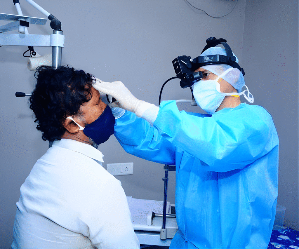
How is Squint Diagnosed?
Eye doctors use advanced diagnostic tools to identify dry eye syndrome.
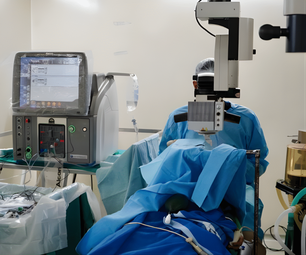
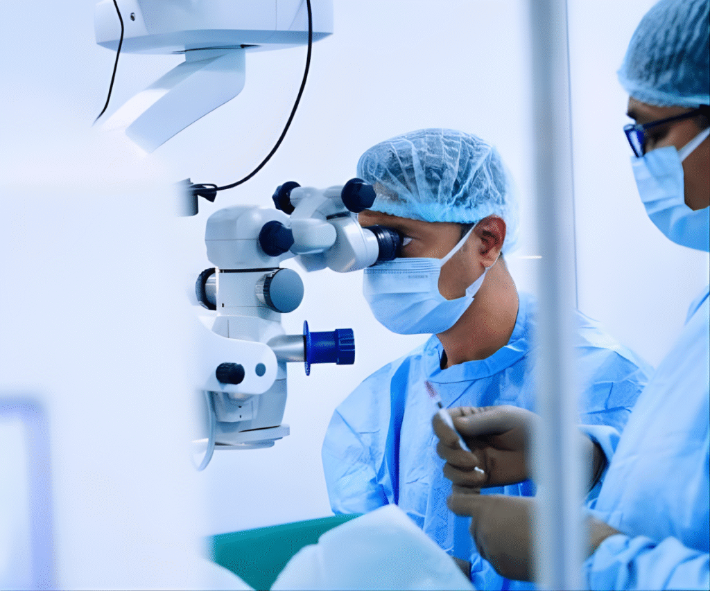
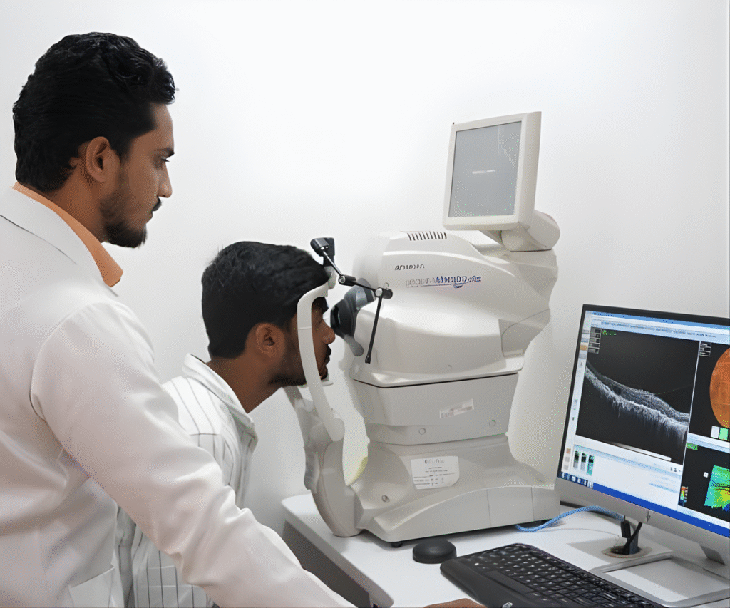
3D Imaging Systems (OCT)
Provides detailed imaging of eye structures to plan accurate surgeries.
Femtosecond Laser Surgery
Enables robotic precision for exceptional outcomes.
Digital Microsurgery Tools
Enables robotic precision for exceptional outcomes.
Eye Alignment Tracking Devices
Enables robotic precision for exceptional outcomes.
Retina Treatments We Offer
Our hospital provides advanced retinal treatments using state-of-the-art technology:
Laser Therapy (Retinal Laser Photocoagulation)
Laser therapy is one of the most commonly performed and highly effective retinal treatments. A carefully focused laser beam is used to treat diseased areas of the retina while protecting healthy tissue
The laser creates tiny controlled burns that seal or destroy abnormal tissue, preventing further damage.
Conditions Treated:
- Retinal Tears or Holes: Laser seals the tear to prevent retinal detachment.
- Diabetic Retinopathy: Laser reduces leakage and stops the growth of abnormal blood vessels.
- Retinal Vein Occlusions: Helps reduce swelling and stabilizes the retina.
Benefits:
- Outpatient procedure
- Quick recovery
- Minimizes risk of future complications
Intravitreal Injections
Intravitreal injections involve delivering medications directly into the eye to target diseases affecting the macula and retina.
This allows high drug concentration exactly where it’s needed.
Common Medications Used:
- Anti-VEGF agents (e.g., Ranibizumab, Aflibercept, Bevacizumab)
- Steroids for reducing inflammation and swelling
Conditions Treated:
- Age-Related Macular Degeneration (AMD): Slows down degeneration and helps preserve central vision.
- Diabetic Macular Edema (DME): Reduces swelling caused by leaky diabetic vessels.
- Retinal Vein Occlusion: Controls edema and prevents vision loss.
Benefits:
- Highly effective for maintaining or improving vision
- Minimal discomfort
- Fast and precise treatment
Vitrectomy Surgery
Vitrectomy is a sophisticated, minimally invasive microsurgery performed using very fine instruments.
The surgeon removes the vitreous gel from inside the eye to access and repair the retina.
Why It’s Done:
- Retinal Detachment Repair
- Macular Hole Closure
- Epiretinal Membrane Removal
- Clearing Vitreous Hemorrhage (bleeding inside the eye)
- Removing Infections or Foreign Bodies
Procedure Highlights:
- Performed using micro-incision techniques
- Often combined with laser, gas or oil tamponade
- Helps restore normal retinal anatomy
Benefits:
- Significant visual improvement in many cases
- Essential for sight-saving in complex conditions
- Faster healing with modern small-gauge systems
Scleral Buckling & Pneumatic Retinopexy
These are specialized surgeries tailored to reattach a detached retina, depending on the type, location, and extent of the detachment.
A silicone band is gently placed around the eyeball to push the eye wall inward, allowing the retina to reattach.
Best For:
- Young patients
- Specific types of tears or breaks
- Longstanding detachments
Advantages:
- Time-tested technique
- Excellent success rate
Each treatment plan is personalized after a detailed retinal evaluation and imaging to ensure the best possible outcome
Retina Treatments We Offer
Our hospital provides advanced retinal treatments using state-of-the-art technology:
Laser Therapy
Targets and seals retinal problem areas while preserving healthy tissue. Commonly used for retinal tears, diabetic retinopathy, and vein occlusions.
Intravitreal Injections
Delivers medications directly into the eye for conditions like macular degeneration, diabetic macular edema, or vein blockages.
Vitrectomy Surgery
Removes the vitreous gel to repair retinal detachments, treat holes, or clear vitreous hemorrhage.
Scleral Buckle & Pneumatic Retinopexy
Specialized surgical techniques to reattach the retina, depending on the type and extent of detachment.
Scleral Buckle & Pneumatic Retinopexy
Specialized surgical techniques to reattach the retina, depending on the type and extent of detachment.
Zeiss VisuMax Laser
Used in combination with other technologies, this laser system is capable of treating complex retinal conditions with precision and minimal side effects.
Each treatment plan is personalized after a detailed retinal evaluation and imaging to ensure the best possible outcome.
FAQ’s
Frequently Asked Question
Floaters, flashes of light, blurry vision, or a dark curtain-like shadow indicate possible retinal damage.
Regular eye exams, especially for diabetics and older adults, help detect issues early and prevent complications.
No, retinal procedures are typically performed under local or mild anesthesia for maximum comfort.
Recovery time varies depending on the procedure but generally ranges from a few days to several weeks.
It depends on the severity of the damage. Early diagnosis and prompt treatment offer the best chance for visual recovery.
Diabetic patients should have yearly checkups, while others should schedule an exam every 1–2 years or as advised by their doctor.
Easy Finance
Pay later with easy finance options
At Dr.Akbar Super Speciality Eye Hospitals, the best eye care hospital in Hyderabad, we offer different treatment plans to make it easier for our patients; by providing a 0% finance* option for all our surgeries, we take a step closer to a holistic patient care policy.
Easy Installments
A refined version of Phaco with even smaller incisions, promoting faster healing and reduced risk of complications.
0% Interest
A refined version of Phaco with even smaller incisions, promoting faster healing and reduced risk of complications.
Instant Approval
A refined version of Phaco with even smaller incisions, promoting faster healing and reduced risk of complications.
Flexible Tenors
A refined version of Phaco with even smaller incisions, promoting faster healing and reduced risk of complications.